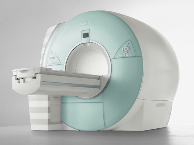
MAGNETIC RESONANCE IMAGING (MR)
Magnetic Resonance Imaging is a medical technique used to identify certain anatomical structures and distinguish the healthy tissues from the diseased ones clearly and accurately by using radio waves in a strong magnetic field generated by large magnets. Unlike the radiograms and computed tomography examinations, MRI does not pose a risk for radiation as no X-rays are used during the procedure.
In our center, SIEMENS MAGNETOM AERA MR device is used.
The tunnel of the device into which the patient enters in has a diameter of 70 cm, which is wider than the conventional MR. In addition, it is very spacious owing to the special lighting. Therefore, the patient is relieved of the fear of entering the tunnel of the device and of the fear of staying at a narrow place. In addition, the Quite Suite (Silent MR) feature provides the patient a more comfortable environment during the procedure. Most of the scanning is performed while the patient’s head is out of the device. In addition, the physician or technician is incessantly in communication with the patient.
Before the procedure, the patient will be asked to put on a gown and take off all metallic items like watches, necklaces, rings, and earrings on herself/himself. Then, the patient lies down on the MR table. The technician will adjust the apparatus helping the patient to keep her/his head stable. The patient will put on headsets, thereby, will be able to communicate with the team in the MR room. She/he will receive the commands from them and act accordingly. The patient will be given a button to inform the MR team if s/he is distressed. Then the patient is taken to the MR device and the shooting process is started.
